Medical device and MedTech insights, news, tips and more
Giant Touchscreen Gives Med Students a 3D View of Human Anatomy
October 12, 2016

 Within weeks of starting medical school, first year students enter the gross anatomy lab at the University of B.C. to begin dissecting a human cadaver.
On this day, 288 students are crowded around 48 bodies donated so they can learn their trade — and that responsibility weighs on would-be doctors like several Earth gravities.
“It’s very exciting and they are nervous,” said instructor and senior radiology resident Kathryn Darras. “It’s a very emotional day.”
This year, for the first time, students will have access to imaging tools that past generations of doctors could only dream of.
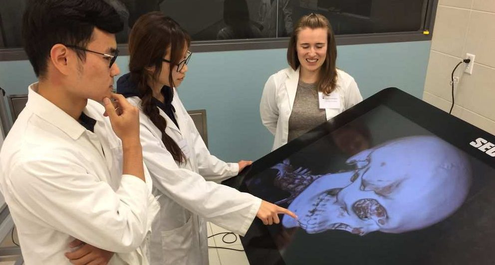
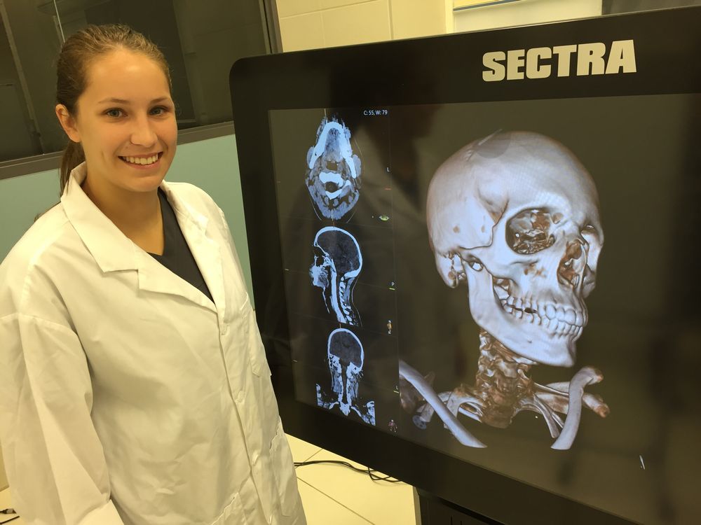
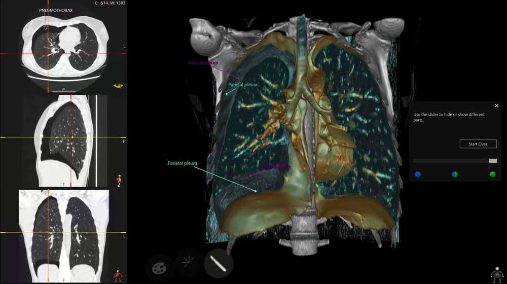
The giant Sectra visualization screen is a powerful three-dimensional imaging system that displays CT (computerized tomography) scans that can be turned, rotated and cut into cross-sections with the stroke of a finger. The students take turns exploring images of body parts they will dissect that day.
“The pressure to do a good job dissecting a donor body is intense,” said first-year medical student Lulu Yang. “It’s a relief to be able to cut into an image of a human being, rather than the real thing.
“When you are cutting into a human body it’s a lot of responsibility, because someone donated this for you. With the Sectra table you can play with it, look at it from all angles and if you make a mistake you can undo.”

The five-foot table — like a massive touch-screen iPad in the corner of the anatomy lab — allows students to see everything from skin, bone and connective tissue to organs and muscles in relation to each other without the disruptive processes of embalming and dissection.The device, the first of its kind in Canada, promises to demystify a complex and life-changing process.
Dissecting all the frogs and rabbits in the world would not begin to prepare you for the prospect of working on a human body, according to student Alison Wookey.
“I took an anatomy class in my undergrad and we dissected bunnies, but it’s nothing like working with a cadaver. It’s totally unbelievable,” she said. “I don’t think I’ll ever see anything the same way again.”
During lab sessions, students gather in small groups around the Sectra screen while Darras shows them structures they will see in the cadavers in unprecedented detail. For example, images of the cervical spine show the shape and orientation of bones in a way that cannot be revealed through dissection.


UBC’s head of radiology chirps in with diagnostic advice when Darras reveals the uppermost vertebra.
“You can see the atlas bone is ring shaped and we know that a ring almost never breaks in just one place. So, if you see one break, don’t stop looking,” said Bruce Forster.
Locally produced images will be loaded on the Sectra in the coming months, after the university and Vancouver Coastal Health work out a patient privacy protocol.
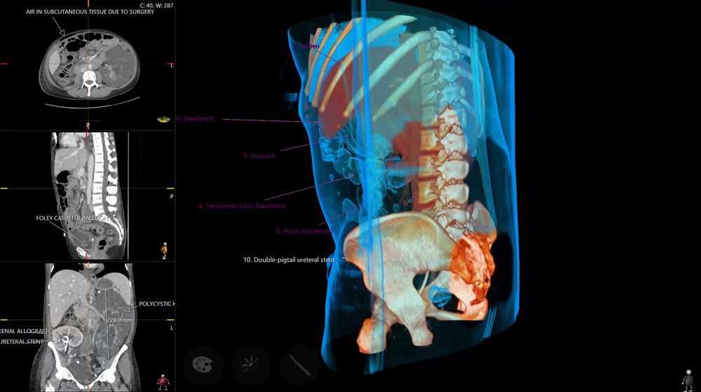
Among the 175 stock images preloaded on the device is a CT scan of a patient after a kidney transplant in which the new kidney is clearly visible from every angle, as well as the apparatus implanted to keep it connected to the bladder, and a cyst-laden kidney intact on the left side of the abdomen.
“Because the patients are alive when these images are taken, you can see how (the kidney) functions in relation to everything around it, the circulatory system and the colon,” said Forster. “If you get an abscess around that kidney you can really appreciate what other structures that will affect.”
The Sectra screen is very much Forster’s baby.
When a colleague sent him a small ad describing the visualization screen four years ago, Forster immediately brought it up with the dean of the medical school, who loved its potential.
Read Full Article – Source: “iPad” imaging tool lets UBC medical students see human anatomy in 3D | Vancouver Sun
Author – Randy Shore
Photo Credit – RANDY SHORE, UBC/PNG
